
Acidic Water Impacts Sea Urchins
Kiel study verifies adverse effects on sea urchin larvae
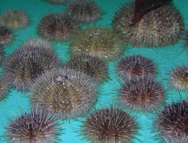
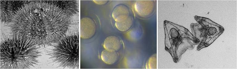
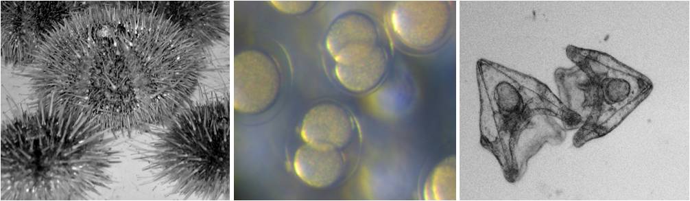
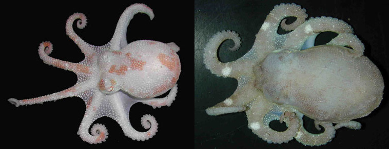
Pluteus larvae of the green sea urchin Strongylocentrotus droebachiensis are tiny, about 0.3 mm long “eating machines” which feed on algae for several weeks in plankton before they go through metamorphosis and begin their life on the sea bed. Thin skeletal elements support their transparent bodies. These spines are made of calcium carbonate and are formed by special cells. Only recently has it been discovered that pluteus larvae grow more slowly in acidified sea water and need more energy. An extended development period, however, increases the risk that the larvae will be eaten by the many predators in the open sea.
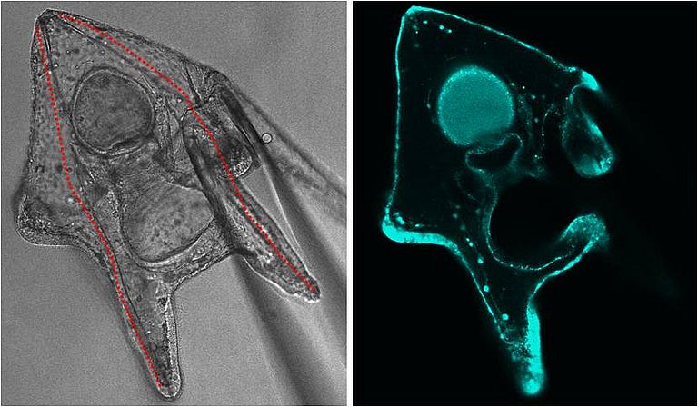
As part of the BMBF coordinated project BIOACID, scientists from GEOMAR Helmholtz Centre for Ocean Research Kiel and the Cluster of Excellence “The Future Ocean” at Kiel University examined which mechanisms lead to this decrease in growth rate. Using methods developed for research on mammals, the scientists were able to show that pluteus larvae cannot control the pH value in their cavities. These measurements were taken with tiny, 2 µm thin pH-electrodes. In contrast, measurements with pH sensitive colorants revealed that the cells in the bodies of larvae can control their internal pH value under the stress of acidification. This is important as the first skeletal elements are formed inside the cell itself – a process that only functions at very uniform conditions. Changes in the pH gradient between the calcifying cells and the cavities could be one of the reasons for the observed reduction in growth and calcification in an acidified sea environment. Under these conditions, more energy is necessary to control the pH value in the calcifying cells. This energy is thus missing for the growth process.
“We need further studies on the cell biology of calcification processes in order to better understand the mechanisms of sensitivity” explains Prof. Dr. Frank Melzner, head of the research group for ecophysiology at GEOMAR. “The pH regulation abilities of tiny larval stages can only be researched with sophisticated optical and electric methods” continues Melzner. “That is why the cooperation with human physiologists at the faculty of medicine is so very important, as they have developed methods over decades that are very useful for us marine biologists.” Prof. Dr. Markus Bleich, director of the Physiological Institute of Kiel University confirms this, “For the first time we can transfer techniques, which we have used successfully on mammals for a long time, to the cell processes of sea urchins. The interdisciplinary research for the coordinated project BIOACID and “The Future Ocean” allows us to examine the disturbances of the inner milieu of the cells of different organisms. Thus we can better understand the physiological processes necessary for vital functions.”
The first co authors of the study, Dr. Meike Stumpp and Dr. Marian Hu, a former PhD student at GEOMAR and a former research assistant at Kiel University, are currently completing one-year postdoc positions at the University of Gothenburg. There they are continuing their research on sea urchin larvae. First results point towards further fundamental findings on the physiology of sea urchin larvae. “These studies show that our understanding of the functioning of early life stages of even the most common sea organisms is still in its infancy – exciting times for interdisciplinary marine research” sums up Prof. Melzner
Original Paper:
Stumpp, M., Hu, M.Y., Melzner, F., Gutowska, M.A., Dorey, N., Himmerkus, N., Holtmann, W., Dupont, S.T., Thorndyke, M.C., Bleich, M., 2012: Acidified seawater impacts sea urchin larvae pH regulatory systems relevant for calcification. Proceedings of the National Academy of Sciences, DOI: 10.1073/pnas.1209174109.
Links:
BMBF coordinated project “Biological Impacts of Ocean ACIDification" (BIOACID), www.bioacid.de
Cluster of Excellence “The Future Ocean”, www.futureocean.org and www.futureocean.org/evolving-ocean
Contact:
Dr. Andreas Villwock, GEOMAR, Communication & Media, Tel.: (0049)431-600-2802, avillwock(at)geomar.de
Friederike Balzereit, Cluster of Excellence “The Future Ocean”, Public Outreach, Tel.: (0049)431-880-3032, fbalzereit(at)uv.uni-kiel.de