

Laborleiter:
Dr. Joachim Schönfeld
Tel.: 0431/600-2315
E-Mail: jschoenfeld(at)geomar.de
Fachkraft für Arbeitssicherheit:
staatl. gepr. Techniker
Dietmar Schmitz
Tel.: 0431/600-2750
E-Mail: dschmitz(at)geomar.de
The micropalaeontology laboratory of the Research Unit Paleoceanography is a facility for micropalaeontological sample preparation. Intention is to allow for a reliable quantification of all remains of organisms within a sediment sample. The laboratory at room 8/D-116 offers all necessary facilities for the preparation of calcareous microfossils (foraminifera, ostracods, pteropods) and the initial preparation for diatom or nannoplankton microscopic slides. Fume hoods in other laboratories are available for the preparation of organic, siliceous, and phosphatic organisms and their remains (dinoflagellates, pollen, diatoms, radiolarians, conodonts, and fish teeth), where aggressive chemicals (acids, lyes) may be applied.
Additional facilities comprise an automated extraction line for biogenic silica in sediments, the TURNER fluorometer for the determination of chlorins (chlorophyll and decay products) within sediments, and a SediGraph for grain size analyses.
In the Micropalaeontology Laboratory, samples are prepared for the study of taxonomy, stratigraphy, ecology, proxy development and application of microfossils. Investigations on recent benthic foraminifera are distribution surveys and temporal sample series studies in combination with environmental observations and hydrographic measurements. Culture and field experiments are pursued as well.
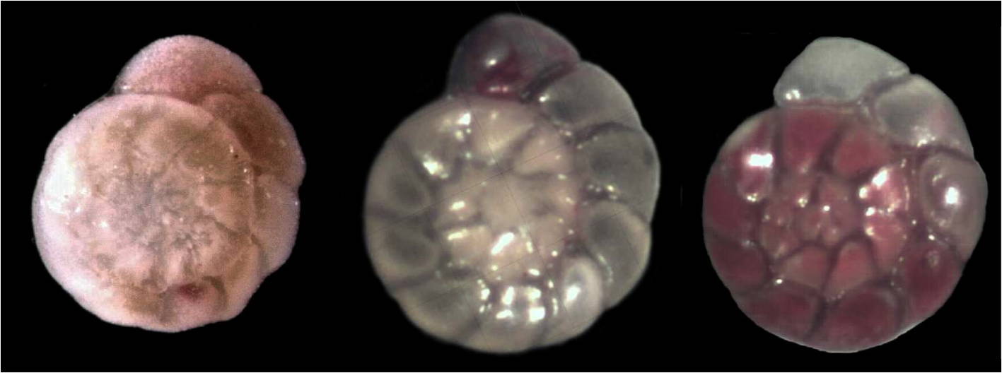
Benthic foraminifera live in the uppermost centimeters of the sediment. Their species composition and population density is in balance with the prevailing environmental conditions. Therefore, many studies focus on living foraminifera. They contain cream, light green, or yellowish-brown cytoplasm. Tests containing cell plasma are difficult to distinguish from specimens that are filled with fine sediment. The plasma can be made visible with specific dyes. We use rose Bengal, which stains the proteins of the cytoplasm. Immediately after sampling, foraminiferal samples are stained and preserved with a solution of 2 grams rose Bengal in one liter ethanol (at least 70%, technical grade, e.g. denatured alcohol).
Foraminiferal assemblages from surface samples also contain empty tests of varying ages. The number of empty tests of the so-called dead assemblage is 10 to 1000 times higher than the number of living individuals. Early diagenetic processes change the composition of the dead fauna over time because the tests of each species have a different preservation potential. When they are deposited in the sediment below the surface layer, the assemblage composition remains constant. The dead assemblage spans many generations from different years, with robust shells of well-preservable species being over-represented. There are also tests of species that have lived in other places, have been transported and deposited at the sampling site.
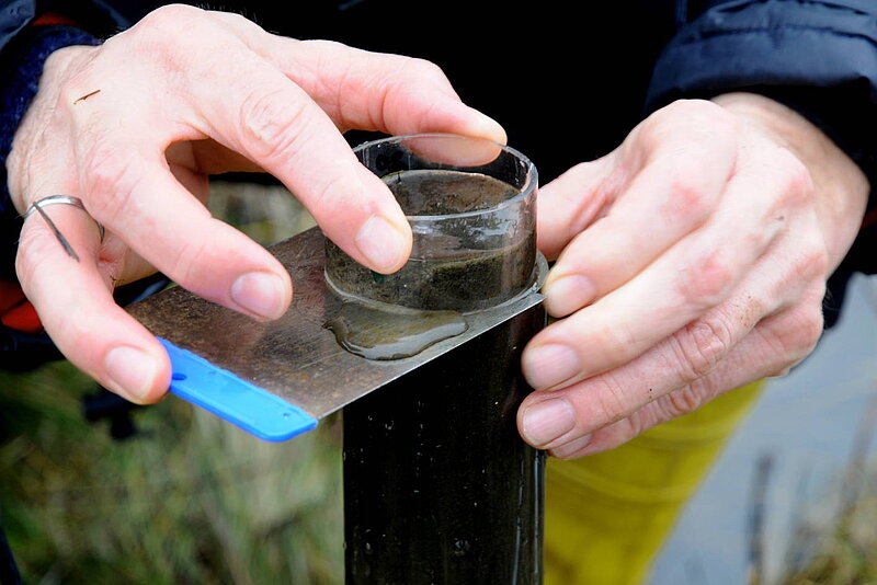
During field work in a given study area, always the same sampling device is used, and the same sample volume is taken. At sea, we use a multicorer or a box corer to recover an undisturbed sediment surface from great water depths. Foraminiferal samples are taken directly from the multicorer cores, or we use hand-held push cores made of stainless steel or polycarbonate with an inner diameter of 36 mm or 54 mm. After recovery, the sediment is pressed out of the multicorer or coring tube until it protrudes by 1 cm. The extruded sediment is stabilized with a polycarbonate ring. The top centimetre is cut off with a shovel spatula, powder spatula or kitchen knife.
A sample taken that way has a volume of 10 or 23 cm3, multicorer tube samples have of volume of 78.5 cm3. The actual sample volume may deviate from the geometric volume. It is therefore necessary to mark the level of the sample fill on the sample vials with a waterproof marker pen before the sample is preserved. The volume is determined in the laboratory after sample washing. Investigations of living foraminifera always refer to the sample volume. Population densities are given in individuals per 10 cm3 of surface sediment. For fossil foraminiferal faunas, abundances refer to the dry weight of the samples. The abundance is given in specimens per gram sediment.
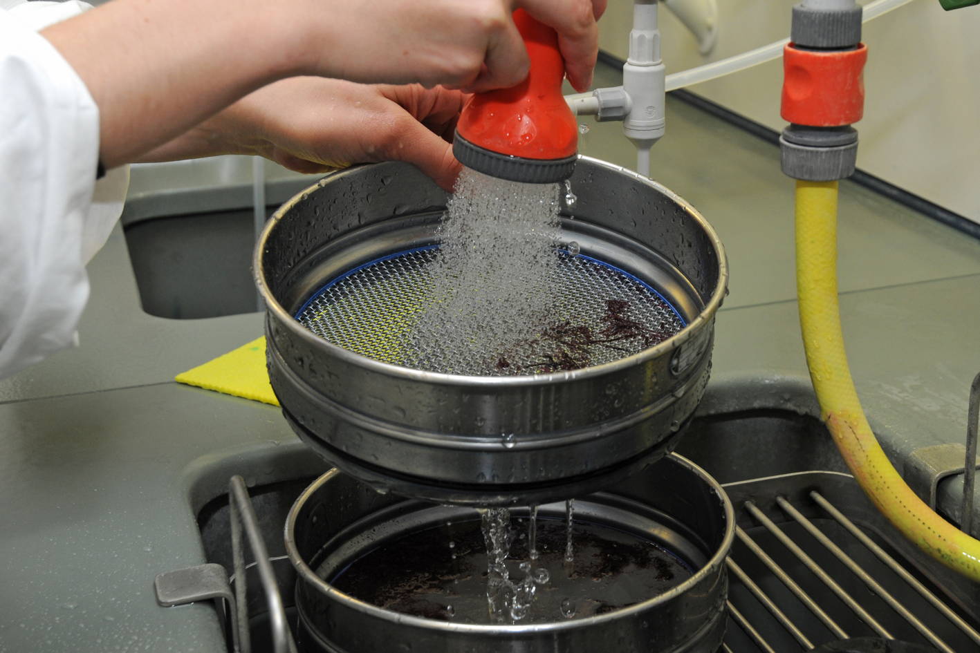
Samples preserved and stained with ethanol have to be stored for at least two weeks to allow a complete staining of individuals that were living at the time of sampling. During the subsequent processing, we first pass the samples through a 2000 µm sieve. This is intended to remove pebbles and other large particles that would grind down fragile foraminiferal tests while washing the sample. The fraction >2000 µm is rinsed on the sieve with a shower, dried at 60°C and stored. Then, the samples are transferred to a 63 µm sieve and the washing continues by using a shower and tap water. It is checked under a microscope from time to time whether the foraminifera are sufficiently clean. After washing, the sample residue is stored under ethanol (at least 70%) if fragile, agglutinated species are to be studied. Otherwise, the 63 – 2000 µm fraction is dried at 60°C, weighed and stored for further investigations.
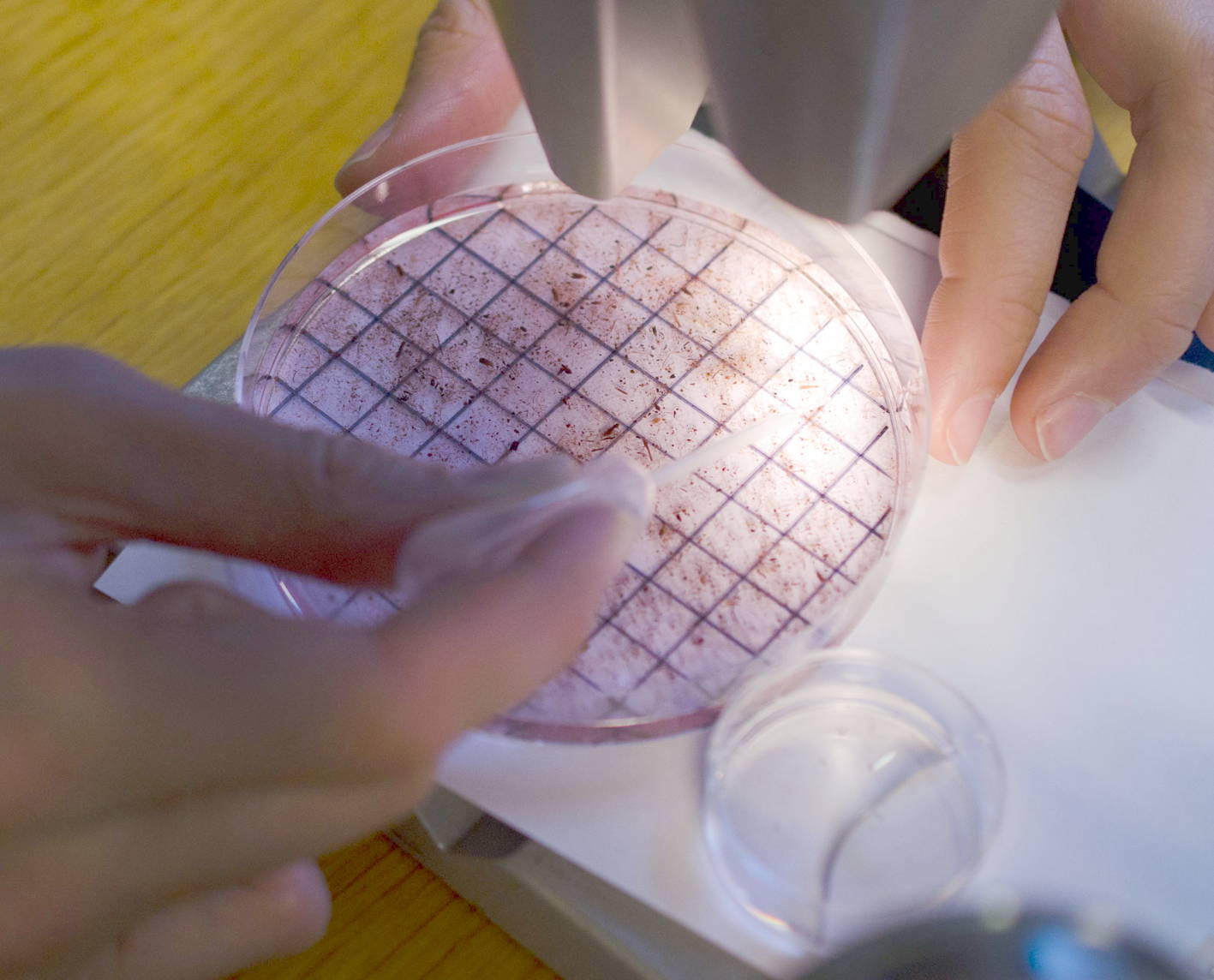
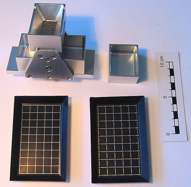
The 63 – 2000 µm fraction is used for taxonomic investigations or distribution studies. The fractions 125 – 2000 µm, 150 – 2000 µm, or, in areas with strong near-bottom currents, the fraction 250 – 2000 µm is used for ecological studies or for proxy development. The coarser fractions are obtained by dry sieved of the fraction 63 – 2000 µm. For picking, a small part of the sample residue is evenly spread on a perforated picking tray. The grains should just be so close together that they do not touch. The picking tray is placed on a counting tray of the same size and examined under the stereomicroscope at 20 to 40 x magnification. The foraminifera are taken up with a lightly greased dissecting needle, moved to a hole and stripped off there. They then fall into the counting tray below. After picking is done, they can be transferred from there to microcells for further investigations.
When working quantitatively, the entire sample or a representative aliquot is picked completely. The assemblage should comprise approximately 200 – 300 specimens if subsequent statistical analyses are intended. If a sample contains substantially more specimens, it will be split accordingly to arrive at the required number of 200 – 300 individuals. An Otto microsplitter is used for dry residues. The splits are weighed. Samples from mud flats or salt marshes must not be dried for splitting. The thin shell walls of agglutinated species collapse once the residue dries. Those species can then no longer be determined. For splitting of wet samples, Scott or Charrieau sedimentation splitters, Motodo or Folsom splitters are used.
To pick wet samples, the residue or the aliquot is transferred to a gridded Petri dish. Foraminifera that have been living at the time of sampling are picked under water cover. Only bright raspberry colored individuals are considered as alive at the time of sampling. The stained specimens are removed with a very thin Pasteur pipette and collected in a small Petri dish. From there they can be transferred with a fine brush (marten hair, grade 000) to microcells or small glass flasks for long-term storage under ethanol or glycerine.
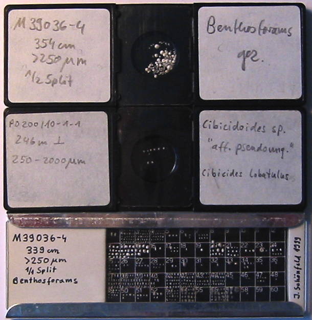
Foraminifera are stored in plastic or cardboard cell slides. The cell slides have a transparent coverslip so that the foraminifera can be viewed with a stereomicroscope. Collective cell slides contain all individuals picked from a sample. They are randomly arranged and not fixed. Only specimens of a specific species are contained in a single cell slide. They are fixed with glue, arranged in rows or sectors so that different sizes or aspects can be compared. Plummer cell slides contain all individuals of a sample. They are sorted by species and fixed with glue. This documents both, the identification of the species and its differentiation from similar taxa, as well as the number of individuals. Before preparing a single cell or Plummer cell slide, the bottom of the cell slide is thinly coated with a water-soluble glue. Wallpaper paste, methylhydroxyethylcellulose or traganth is used. When the glue has dried, it forms an invisible film. Foraminifera are placed and arranged on this film with a very fine, wet paint brush (marten hair, grade 000). The moisture makes the glue film swell up a bit. The foraminifera sink into the gel layer. When the moisture has evaporated, the foraminifera are fixed. They can be removed again with a wet paint brush without leaving any residue, e.g. to be placed elsewhere in the cell, or to be removed for chemical analysis or imaging with a scanning electron microscope.
All specimens of a species are arranged in rows in one or more adjacent fields in the Plummer cell slide. They are aligned with the youngest chamber at the top, and then fixed. We start with the largest specimens of the species and add the next smaller individuals. This makes it easier to discriminate a species. Even the smallest individuals can be assigned with certainty at the end. The species are then determined and the individuals are counted.
Station protocol for sampling at sea
Station protocol example
(Data published in Cruise Report ALKOR 438 [AL438] - Foraminiferal biomonitoring in the North Sea, May 29 - May 31, 2014, Kiel (Germany) - Kiel (Germany). -- GEOMAR Helmholtz-Zentrum für Ozeanforschung, Kiel, 54 pp., doi 10.3289/CR_AL438.)
Counting form for Plummer cell slides
Counting form example
(Data published by Schönfeld, J. & Mendes, I., 2021.Environmental triggers of faunal changes revealed by benthic foraminiferal monitoring. -- Estuarine, Coastal and Shelf Science, 253, 107313, 16 pp., doi: 10.1016/j.ecss.2021.107313.)